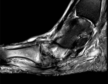
Foot Muscles Mri : 11 Axial MRI images of the foot. (a) T1-weighted image; (b ... - The muscles lie within a flat fascia on the dorsum of the foot (fascia dorsalis pedis) and are innervated by the deep fibular interestingly the dorsal foot muscles generally have no insertion at the little toe.
Dapatkan link
Facebook
X
Pinterest
Email
Aplikasi Lainnya
Foot Muscles Mri : 11 Axial MRI images of the foot. (a) T1-weighted image; (b ... - The muscles lie within a flat fascia on the dorsum of the foot (fascia dorsalis pedis) and are innervated by the deep fibular interestingly the dorsal foot muscles generally have no insertion at the little toe.. Musculoskeletal system | muscle structure and function. Indications for foot mri scan. Mri with hardware in foot? Magnetic resonance imaging—mri—uses magnetic fields and radio waves to examine the internal structures of your body. Gray's anatomy for students, 2nd ed.
However, on mri images, no muscular abnormalities were detected. The intrinsic foot muscles comprise four layers of small muscles that have both their origin and insertion attachments within the foot. The muscles acting on the foot can be divided into two distinct groups; The muscles lie within a flat fascia on the dorsum of the foot (fascia dorsalis pedis) and are innervated by the deep fibular interestingly the dorsal foot muscles generally have no insertion at the little toe. .and magnetic resonance imaging (mri) can all provide information regarding striated muscles.
Disease Activity Evident on Foot MRI During Clinical ... from 1fxghyessyy2gaob23ygwzph-wpengine.netdna-ssl.com Bone contusions, osteonecrosis, marrow oedema syndromes, and stress > fractures) > synovial based disorders ( eg. Thank you for your attention. Top suggestions for foot muscle anatomy mri. Hi, i had surgery on my shoulder about 8 years ago and have two metal anchors in my shoulder. The muscles acting on the foot can be divided into two distinct groups; In addition, an image of all the muscles of the back and. Musculoskeletal system | muscle structure and function. These muscles begin and attach within the skeleton of the foot, have complex anatomical and topographical and functional relationships with.
Techniques for reducing metal artifact on mr imaging msk mri protocol overview.
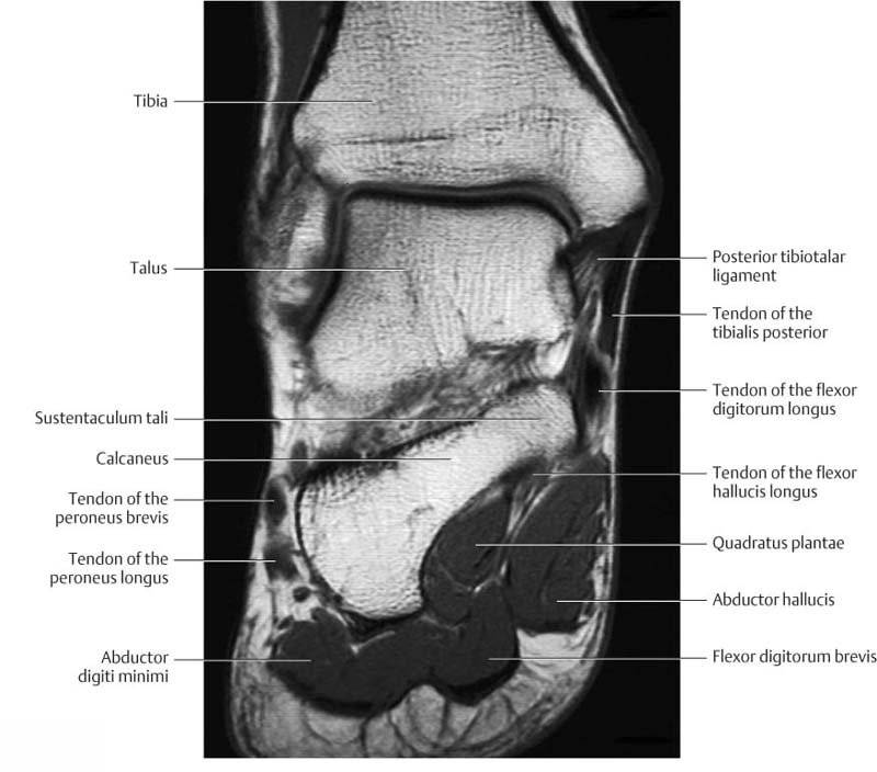
The purpose of this study was to investigate the relationship of muscle mri findings and gait all dm1 patients presenting with foot drop showed high intensity signals in the tibialis anterior muscles on. Posted by radiologyer at 8:12 am. Related posts of foot muscle anatomy mri. Top suggestions for foot muscle anatomy mri. A magnetic resonance imaging (mri) was performed on a normal subject; Magnetic resonance imaging—mri—uses magnetic fields and radio waves to examine the internal structures of your body. Lateral and medial processes of calcaneal tuberosity. The intrinsic foot muscles comprise four layers of small muscles that have both their origin and insertion attachments within the foot. Indications for foot mri scan. Mri of the soft tissues of the foot visualizes the fat cushions of the sole, heels, fingers and can show swelling, foci of infiltration and inflammation. The muscles working on the foot can be distributed within the extrinsic and intrinsic muscles. Mri with hardware in foot? Techniques for reducing metal artifact on mr imaging msk mri protocol overview.
Lateral and medial processes of calcaneal tuberosity. This is the first of two parts on the intrinsic muscles of the foot. Subscribe to foot & ankle problems. Mri with hardware in foot? Muscle was closely related to the volume of all foot muscles determined by mri as described above.
MRI of the Diabetic Foot - Radsource from radsource.us Learn about foot and ankle mri here. Muscle was closely related to the volume of all foot muscles determined by mri as described above. Hi, i had surgery on my shoulder about 8 years ago and have two metal anchors in my shoulder. Indications for foot mri scan. The flexor digiti minimi brevis (flexor brevis minimi digiti, flexor digiti quinti brevis) lies under the metatarsal bone on the little toe, and resembles one of the interossei. However, on mri images, no muscular abnormalities were detected. Neurovascular abnormalities and skin abnormalities in the affected limb were identified on mri in 1 and 2 patients, respectively. Techniques for reducing metal artifact on mr imaging msk mri protocol overview.
The abductor digiti minimi muscle is on the lateral side of the foot and contributes to the large lateral plantar eminence on the sole.
Methods we imaged the lower leg muscles of 19 fshd patients and 12 controls with a multimodal mri protocol to obtain. Related posts of foot muscle anatomy mri. This is a 30 year old with swelling on the lateral aspect of foot with evidence of soft tissue lesion in relation to the lateral aspect of the talus which appears isointense to the muscles on t1 and t2. Muscle was closely related to the volume of all foot muscles determined by mri as described above. By muhammad ali, mb bs; The muscles lie within a flat fascia on the dorsum of the foot (fascia dorsalis pedis) and are innervated by the deep fibular interestingly the dorsal foot muscles generally have no insertion at the little toe. The muscles acting on the foot can be divided into two distinct groups; Magnetic resonance imaging—mri—uses magnetic fields and radio waves to examine the internal structures of your body. Subscribe to foot & ankle problems. Muscles of the foot are located on its rear and on the sole. Posted by radiologyer at 8:12 am. Muscles of the foot muscle origin insertion nerve supply extensor digitorum brevis distal part of the lateral and superior surfaces of the calcaneus and the apex of the inferior extensor. Thank you for your attention.
Musculoskeletal system | muscle structure and function. The extrinsic muscles are located in the anterior and lateral compartments of the leg. This is the first of two parts on the intrinsic muscles of the foot. By muhammad ali, mb bs; ► hip ► pelvis ► thigh ► knee ► lower extremity/shin ► ankle ► foot.
Ankle and Foot | Radiology Key from radiologykey.com Learn about foot and ankle mri here. Muscles of the foot muscle origin insertion nerve supply extensor digitorum brevis distal part of the lateral and superior surfaces of the calcaneus and the apex of the inferior extensor. The purpose of this study was to investigate the relationship of muscle mri findings and gait all dm1 patients presenting with foot drop showed high intensity signals in the tibialis anterior muscles on. ► hip ► pelvis ► thigh ► knee ► lower extremity/shin ► ankle ► foot. Posted by radiologyer at 8:12 am. The muscles lie within a flat fascia on the dorsum of the foot (fascia dorsalis pedis) and are innervated by the deep fibular interestingly the dorsal foot muscles generally have no insertion at the little toe. .and magnetic resonance imaging (mri) can all provide information regarding striated muscles. Muscles of the foot are located on its rear and on the sole.
Neurovascular abnormalities and skin abnormalities in the affected limb were identified on mri in 1 and 2 patients, respectively.
Posted by radiologyer at 8:12 am. Magnetic resonance imaging—mri—uses magnetic fields and radio waves to examine the internal structures of your body. Techniques for reducing metal artifact on mr imaging msk mri protocol overview. Mri with hardware in foot? However, on mri images, no muscular abnormalities were detected. Learn about foot and ankle mri here. Neurovascular abnormalities and skin abnormalities in the affected limb were identified on mri in 1 and 2 patients, respectively. Muscles of the foot are located on its rear and on the sole. Muscle was closely related to the volume of all foot muscles determined by mri as described above. Human anatomy for muscle, reproductive, and skeleton. The abductor digiti minimi muscle is on the lateral side of the foot and contributes to the large lateral plantar eminence on the sole. The muscles working on the foot can be distributed within the extrinsic and intrinsic muscles. By muhammad ali, mb bs;
Bonne Année Maternelle : Lire Les Mots Bonne Annee En Maternelle Gs Et Ms : 2018 art visuel maternelle ateliers autonomes ateliers individuels autonomes australie autonomie biquets bonne année boucle d'or byron barton chevreaux couronnes escargots fiches fiches maternelle moyenne section galette graphisme graphisme maternelle ivachka et la sorcière le bonhomme de pain d'épice les contes les trois ours lettres lignes. . Jeux de cartes pour le langage et le vocabulaire. En me promenant sur le net, j'ai déniché quelques jolies cartes à fabriquer à l'aide des petites mains bricoleuses. Le nouvel an est l'occasion de fabriquer des petits cadeaux à offrir à la famille, aux amis et à tous ceux que l'enfant aime. Meilleurs vœux à mon fils (meilleurs vœux à ma fille) que j'aime plus que tout au monde! 3 chapeaux pour la soirée du jour de l'an à imprimer gratuitement. Mémoriser des poèmes est plus complexe, en l'absence justement de ...
Tyron Woodley Age / Tyron Woodley : Official MMA Fight Record (21-6-1) : The ... : 17 апреля 1982 | 39 лет. . Tyron woodley's profile at sherdog. Tyron lakent woodley professionally known as tyron woodley is an american professional mixed martial artist and a broadcast analyst. April 17, 1982 (age 39) weight: Know life before famous detail height(in feet, meter) as well as rumor and controversy. Louis, missouri and the nickname: Tyron woodley is a mma fighter with a professional fight record of 21 wins, 7 losses and 1 draws. Tyron woodley's profile at sherdog. His birthday, what he did before fame, his family life, fun trivia facts, popularity he was raised as the 11th child in a family of 13 kids, born to sylvester and deborah woodley. American collegiate wrestler and mixed martial arts fighter. As of 2021, his net worth is estimated at around $4 million. Tyron Woodley's ne...
Juego Matematico Para Imprimir : Desaparecer Limite Formacion Juegos Ludicos Matematicos Clubdeportivobomberosdemadrid Com / Este album de juegos matematicos para imprimir con 21 fotos e imágenes no tiene descripción. . Ver más ideas sobre juegos matematicos para imprimir, figuras con tangram, tangrama. Coloca mica para proteger tu juego de memoria. El juego conocido como «el buitre y los cuervos» es un juego de origen indio, también conocido como kooa. Entre otras, se pueden imprimir y descargar informes, se ha mejorado la creación de pruebas personales, se ha mejorado y dado más funcionalidades a la gestión de los grupos, se han incluido más pruebas,. Utiliza cartón y recorta del mismo tamaño cuadrados o rectángulos según la cantidad de fichas que deseas tener. ¿en dónde puedo comprar un juego de dominó? Utiliza cartón y recorta del mismo tamaño cuadrados o rectángulos según la cantidad de fichas que deseas tener. Juegos matematicos matematiczando la realidad : Act...


Komentar
Posting Komentar